Stake attention in this memory
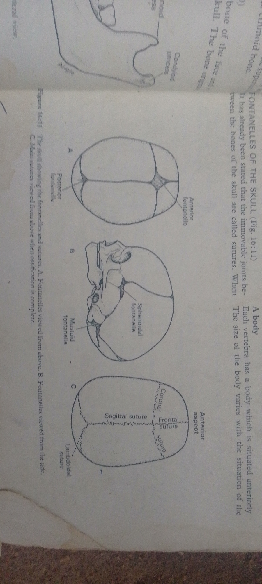
The image displays anatomical diagrams of a baby's skull, illustrating fontanelles and sutures. The diagrams are labeled A, B, and C, with A showing a superior view, B a lateral view, and C a view after ossification is complete. Various fontanelles, including the anterior, posterior, and sphenoidal fontanelles, are indicated. Sutures such as the sagittal, coronal, frontal, and lambdoidal sutures are also clearly marked. Accompanying text explains that fontanelles are joints between skull bones and that sutures are immovable joints. The diagrams are black and white line drawings, typical of educational or medical textbooks. The text also mentions "Figure 16:11" and describes the views presented. The overall presentation is informative and educational, focusing on the anatomical structure of the infant skull.
No transactions found