Stake attention in this memory
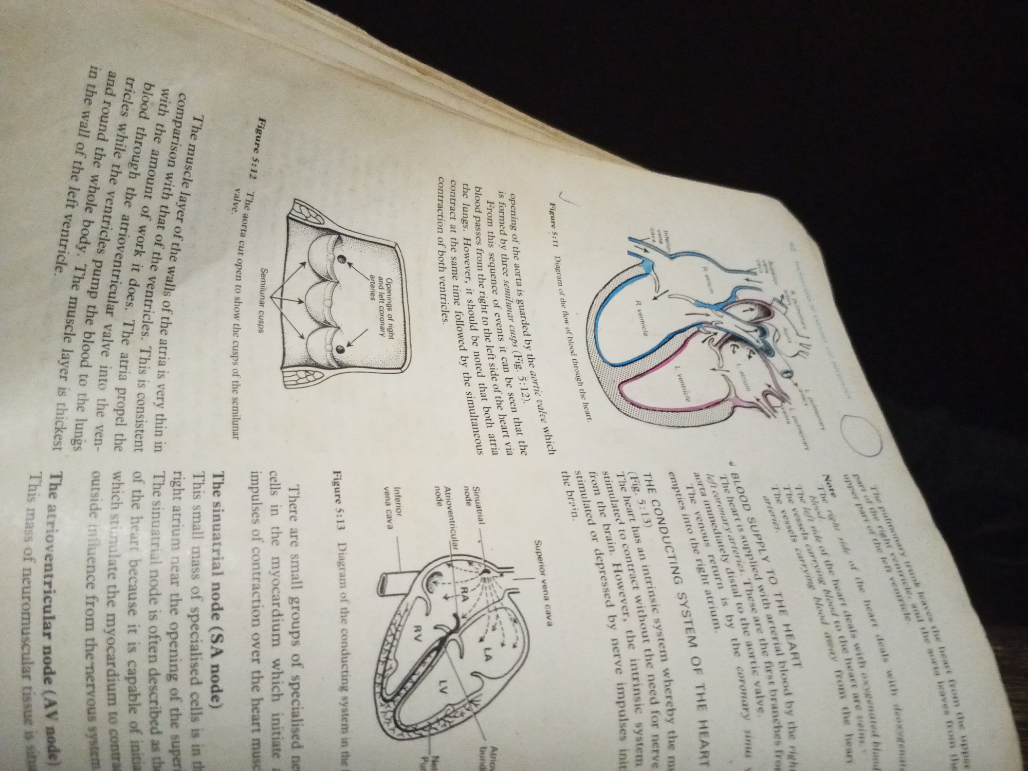
The image displays a page from a biology textbook, open to a section discussing the heart. Two diagrams of the heart are visible. The first diagram, labeled Figure 5:12, shows the aorta with semilunar cusps. The second diagram, labeled Figure 5:13, illustrates the conducting system of the heart, including the Sinuatrial (SA) node, Atrioventricular (AV) node, and the atria (RA, LA) and ventricles (RV, LV). Accompanying text describes the function of the heart's muscle layer and its conduction system. The image is taken from a top-down perspective, and the lighting suggests an indoor environment, likely a study or library. There are no people or any indication of a city or specific location within a city.
No transactions found