Stake attention in this memory
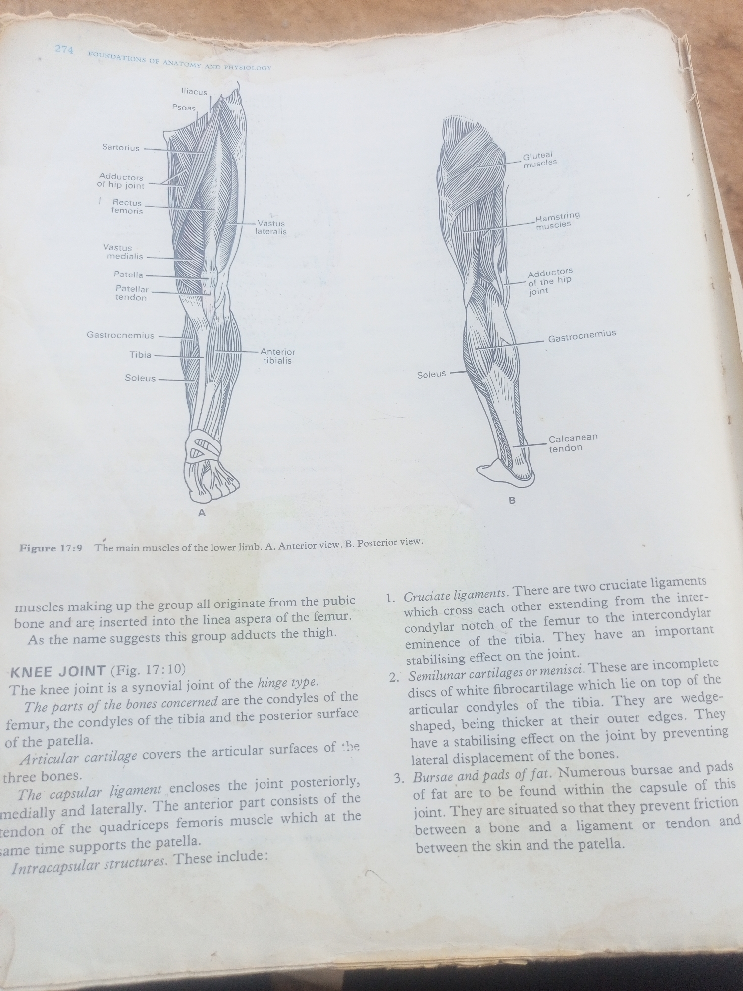
This media file is a photograph of an open book showing anatomical illustrations of the human leg muscles and a detailed description of the knee joint. The left side of the image displays an anterior view (A) of the muscles of the lower limb, with labels such as Iliacus, Psoas, Sartorius, Rectus femoris, Vastus medialis, Patella, Patellar tendon, Gastrocnemius, Tibia, and Soleus. The right side presents a posterior view (B) of the muscles, featuring labels like Gluteal muscles, Hamstring muscles, Adductors of the hip joint, Gastrocnemius, and Calcanean tendon. Below the illustrations, a caption reads "Figure 17:9 The main muscles of the lower limb. A. Anterior view. B. Posterior view." The text below the figure describes the adductor muscles of the thigh and then delves into the anatomy and structures of the knee joint, including its type, the bones involved, articular cartilage, capsular ligament, and intracapsular structures such as cruciate ligaments, semilunar cartilages (menisci), and bursae and pads of fat. The image is a page from a textbook, indicated by the page number "274" and the title "FOUNDATIONS OF ANATOMY AND PHYSIOLOGY" at the top. The paper appears slightly aged, with a textured edge at the top. The lighting suggests an indoor setting, likely a library or study area. No people or specific events are depicted; the focus is purely on the educational content of the textbook. There are no specific time of day, weather, or emotional cues evident. The location context of Jalingo, Nigeria, is not discernible from the image itself.
Symbol
60B26
Volume
11,550
Creator
+$0.23
Revenue
+$0.48
TVL
$18.00
2
Rev Bot 🤖💰
Injected revenue 1h ago
“Revenue bonus for the last stake.”
+$0.53 USD