Stake attention in this memory
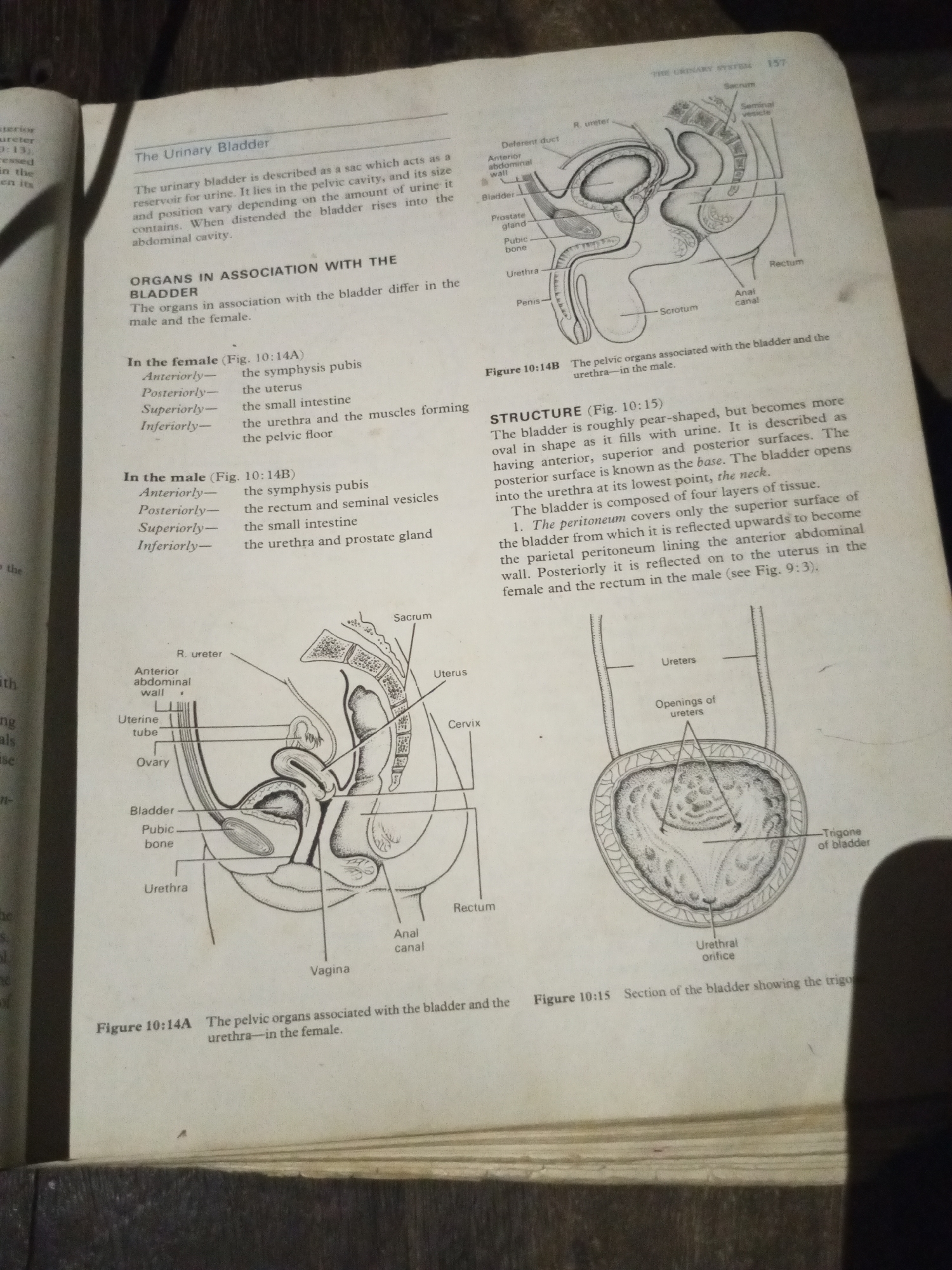
The image displays a page from an anatomy textbook, detailing the urinary system. The page is divided into text and anatomical illustrations. The text describes the urinary bladder, its function as a reservoir for urine, and its changing shape and position based on the amount of urine it contains. It also details the organs associated with the bladder in both females and males, listing them anteriorly, posteriorly, superiorly, and inferiorly. The text further explains the structure of the bladder, its layers of tissue, and peritoneal covering. There are three primary illustrations: Figure 10:14B shows a sagittal view of the male pelvic organs, illustrating the bladder, prostate gland, seminal vesicle, urethra, penis, rectum, and surrounding structures like the pubic bone and scrotum. Figure 10:14A presents a sagittal view of the female pelvic organs, depicting the bladder, pubic bone, urethra, vagina, uterus, cervix, ovaries, and uterine tube, along with the anterior abdominal wall and rectum. Figure 10:15 provides a superior view of the bladder with a section removed, showing the openings of the ureters and the trigone of the bladder, and the urethral orifice. The illustrations are line drawings, typical of educational materials, with labels pointing to specific anatomical parts. The page appears to be from a book lying open, with the binding visible along the left side of the image. The background is dark, suggesting the image was taken in a dimly lit environment, possibly a room with low ambient light. There are no people, actions, or interactions depicted in the image; it is purely an informational diagram.
Symbol
AB87B
Volume
8,265
Creator
+$0.09
Revenue
+$0.16
TVL
$7.42
2
Rev Bot 🤖💰
Injected revenue 4m ago
“Revenue bonus for the last stake.”
+$0.18 USD