Stake attention in this memory
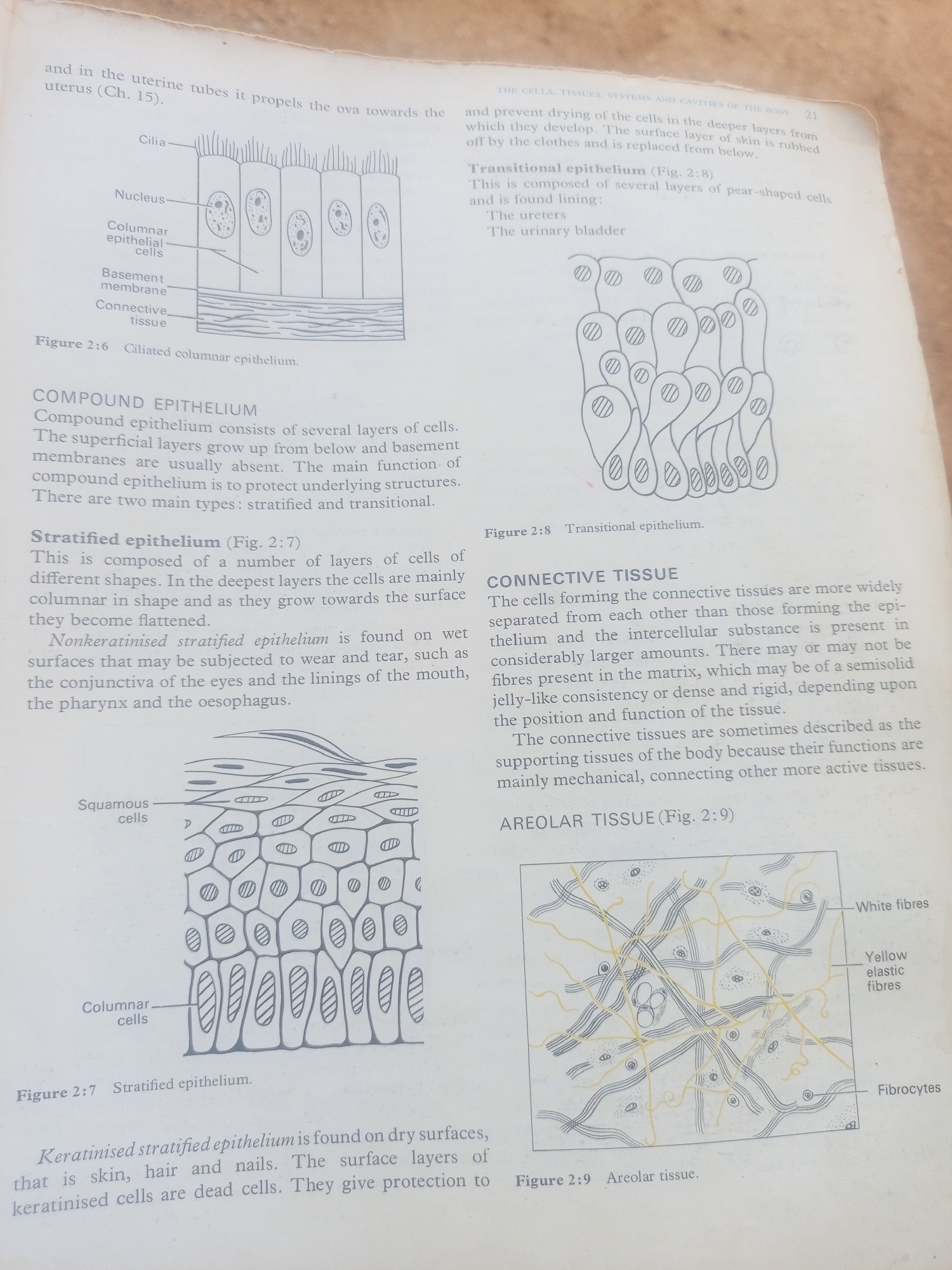
The image displays two pages from a biology textbook, featuring diagrams and text related to epithelial and connective tissues. The content is educational, with a focus on histology. The left page has a diagram labeled "Figure 2:6 Ciliated columnar epithelium," illustrating the structure of these cells with labels for cilia, nucleus, columnar epithelial cells, basement membrane, and connective tissue. Below this is a section defining "COMPOUND EPITHELIUM" and introducing its two main types: stratified and transitional. Further down, "Stratified epithelium (Fig. 2:7)" is described and illustrated with a diagram showing multiple layers of cells, including squamous cells on the surface and columnar cells deeper within. The text explains non-keratinised stratified epithelium found on wet surfaces. The right page begins with text discussing the "Transitional epithelium (Fig. 2:8)," noting it is found lining the ureters and urinary bladder, accompanied by a diagram of this tissue type. The main heading on this page is "CONNECTIVE TISSUE," with a detailed description of its cellular structure, intercellular substance, and fibers. It explains the supporting role of connective tissues. Finally, "AREOLAR TISSUE (Fig. 2:9)" is introduced with a diagram illustrating white fibers, yellow elastic fibers, and fibrocytes. Visible text includes chapter references like "(Ch. 15)," page numbers such as "21," figure labels ("Figure 2:6," "Figure 2:7," "Figure 2:8," "Figure 2:9"), and labels within the diagrams ("Cilia," "Nucleus," "Columnar epithelial cells," "Basement membrane," "Connective tissue," "Squamous cells," "Columnar cells," "White fibres," "Yellow elastic fibres," and "Fibrocytes"). The overall tone is academic and informative. There are no people, specific settings, or indications of time of day or weather; the focus is solely on the scientific illustrations and explanations.
No transactions found