Stake attention in this memory
clinical
analytical
educational
formal
precise
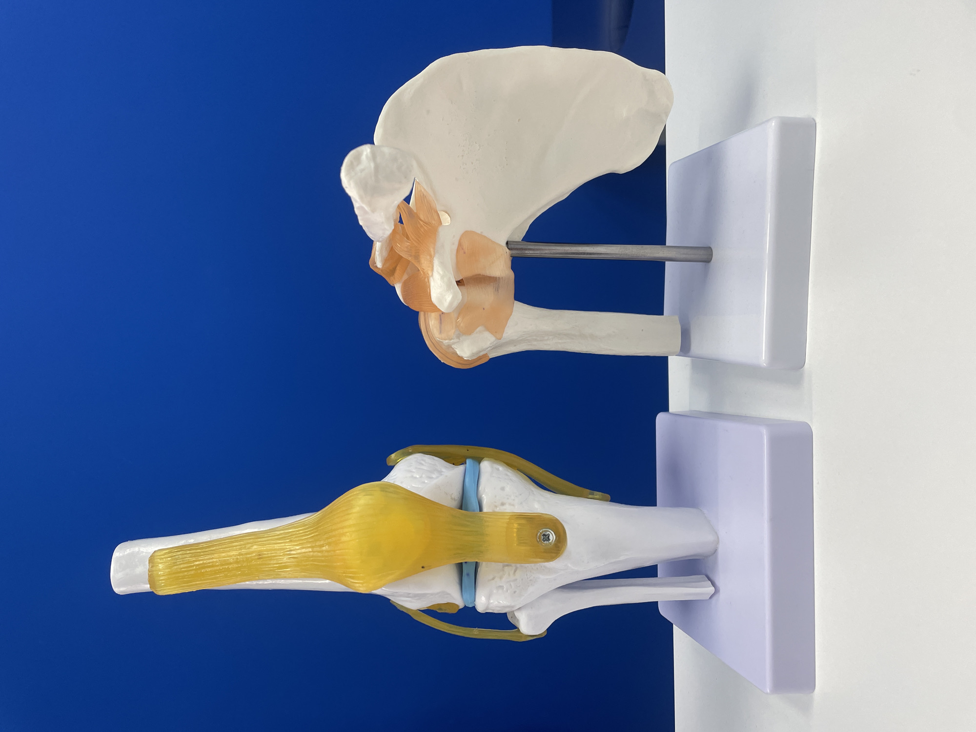
The image displays two anatomical models of human joints: a knee joint and a shoulder joint. The knee joint model, on the left, is white and yellow, showcasing the femur, tibia, patella, and ligaments. The shoulder joint model, on the right, is white and orange, depicting the scapula, humerus, and associated muscles and ligaments. Both models are placed on white bases against a solid blue background. The scene appears to be set up for educational purposes. There is no discernible location in Bishkek City, Kyrgyzstan, or any people present.
transactions
revenues
stakers
Earliest
Latest
Highest stake
No transactions found