Stake attention in this memory
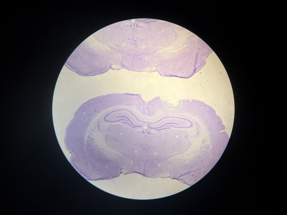
This image presents a microscopic view of two distinct, irregularly shaped slices of biological tissue, consistent with mammalian brain matter, prepared and stained for histological examination. The tissue sections display a predominant purplish coloration, indicative of a hematoxylin-based stain, with variations in cellular density appearing as differential shading. The left tissue slice prominently features identifiable structures resembling hippocampal formations, including the characteristic curvilinear profiles of the dentate gyrus and cornu ammonis (CA) subfields, surrounded by cortical layers. The right tissue slice shows a cross-section of cerebral cortex with similar cellular organization. Both tissue fragments exhibit uneven, somewhat ragged edges, suggesting microtome sectioning artifacts. The entire scene is captured within a circular field of view, characteristic of an optical microscope's eyepiece, set against a uniform black background. The location of this observation is Mexico City, Mexico.
Symbol
044C0
Volume
1,148
Creator
+$0.00
Revenue
+$0.00
TVL
$0.45