
Sign in
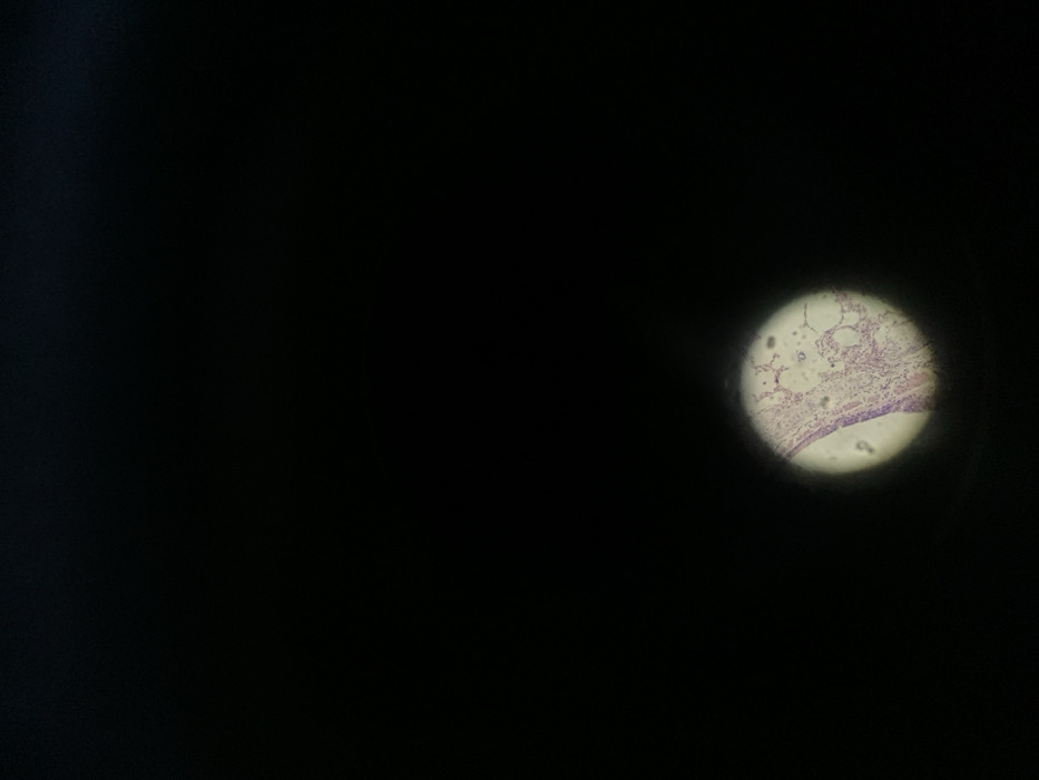
A microscopic view reveals lung tissue, stained with a pink dye, visible within a circular field. Alveoli, air sacs responsible for gas exchange, appear as bubble-like structures. A thin layer of cells, probably epithelial cells, lines the lower edge of the field. The surrounding area is dark and out of focus, creating a tunnel vision effect on the lung cells inside the visible bright area.
Emma🇨🇳💕

Nov 24, 2025, 2:04 AM

Shenyang City, China

Stake attention in this memory

microscope image
histology
lung tissue
stained
cellular structure

A microscopic view reveals lung tissue, stained with a pink dye, visible within a circular field. Alveoli, air sacs responsible for gas exchange, appear as bubble-like structures. A thin layer of cells, probably epithelial cells, lines the lower edge of the field. The surrounding area is dark and out of focus, creating a tunnel vision effect on the lung cells inside the visible bright area.

Loading AttnAds…

transactions
revenues
stakers
Earliest
Latest
Highest stake

No transactions found
More from this user

More from Shenyang City

Emma🇨🇳💕

Nov 24, 2025, 2:04 AM

Shenyang City, China

Licensed under Creative Commons Attribution 4.0 International License. You are free to share and adapt this content with proper attribution.