
Stake attention in this memory
clinical
diagnostic
medical
laboratory
detailed
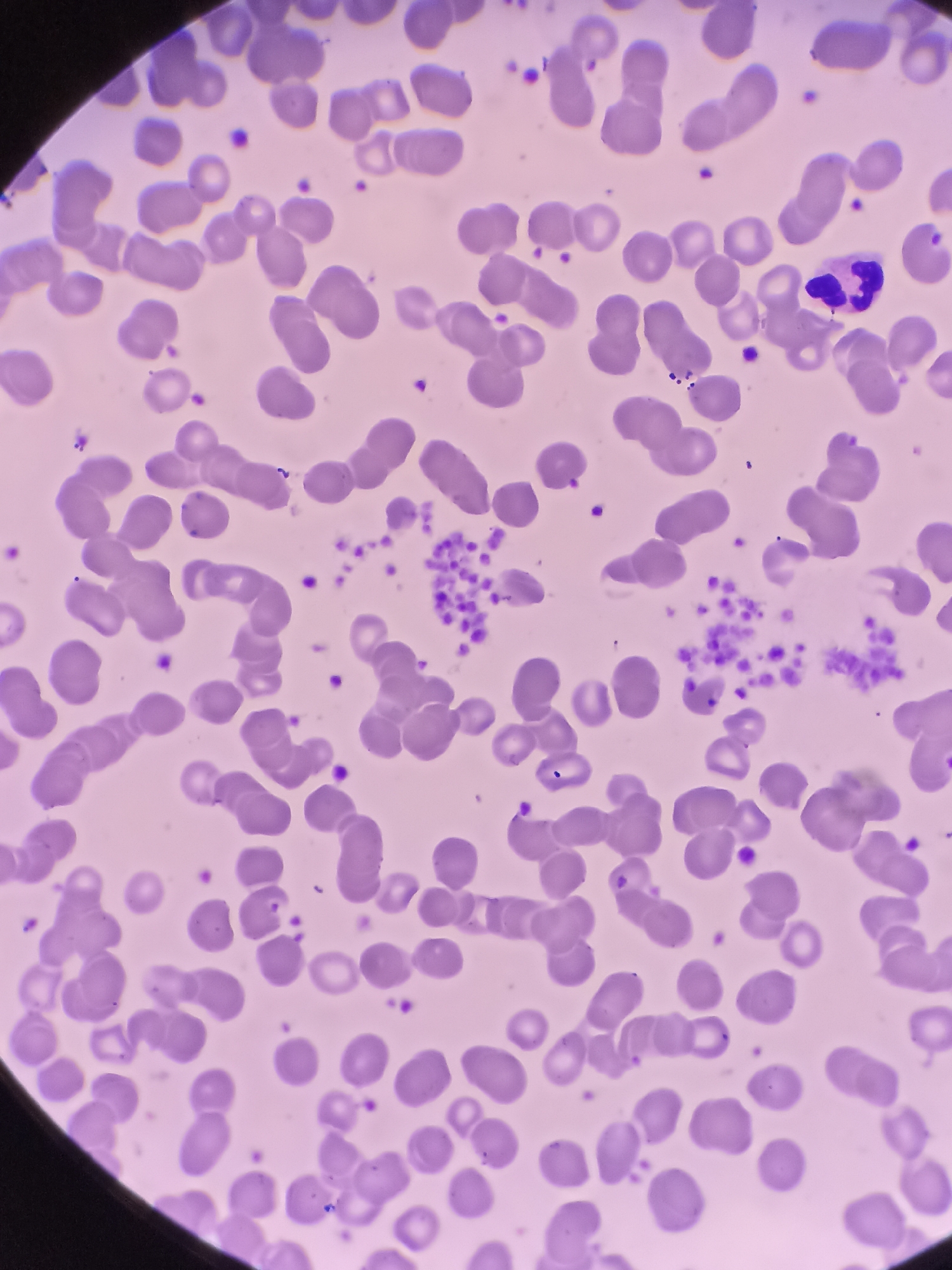
The image is a microscopic view of a blood smear. It displays numerous red blood cells, which are biconcave discs and appear purplish in color due to staining. Scattered among the red blood cells are smaller, irregularly shaped fragments, which are platelets. Some of these platelets are clumped together, forming aggregates. There is also a single white blood cell visible, characterized by its lobed nucleus and granular cytoplasm. The background is a uniform pale pink, indicating the presence of plasma or the staining medium. The image does not contain any elements that would allow for the identification of a location in Surabaya, Indonesia, or any forensic details.
Loading AttnAds…
transactions
revenues
stakers
Earliest
Latest
Highest stake
No transactions found